Breast Cancer Lymph Node Color Doppler Ultrasound Color Meanings

Normal Lymph Node On Ultrasound Lymph Nodes Typically Are Smooth Download Scientific Diagram
Color Doppler Sonography Characterizing Breast Lesions

Typical Us Appearance Of Lymph Nodes Typical Reactive Node Image A Download Scientific Diagram
Reactive Vs Malignant Lymph Nodes Ultrasound Features Radiology Reference Article Radiopaedia Org

Pdf Ultrasonography Of Superficial Lymph Nodes Benign Vs Malignant
Ultrasound Evaluation Of Regional Lymph Nodes As An Extension Of The Breast Ultrasound Exam Youtube
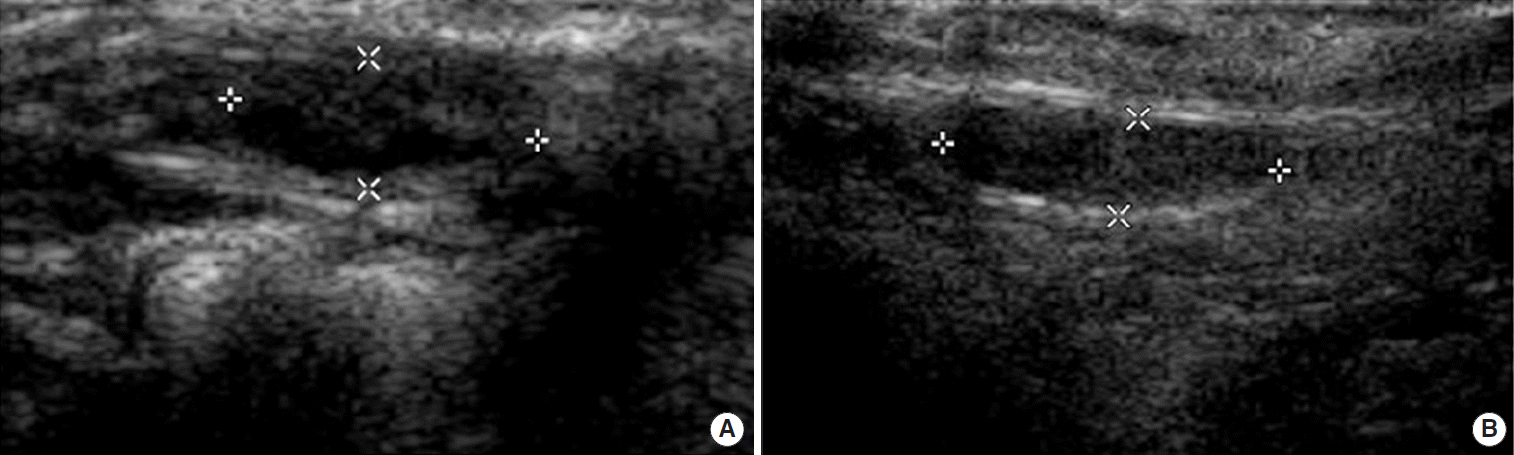
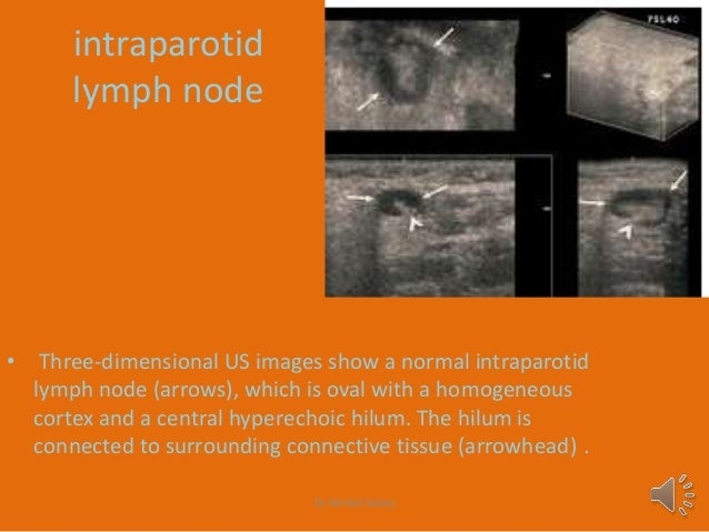
B d three dimensional multiplanar reformatted images also show loss of the fatty hilum.
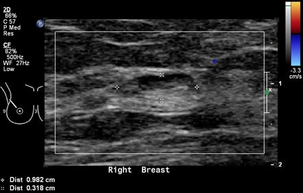
Breast cancer lymph node color doppler ultrasound color meanings. Axillary lymph node dissection alnd is the definitive method to diagnose axillary metastasis but sentinel lymph node biopsy slnb has supplanted this procedure as the primary method of evaluating the axilla in most cases of early stage breast cancer because slnb has a significantly lower rate of morbidity than does alnd and a low false. The presence of cancer cells is known as lymph node involvement. Lymph nodes are small bean shaped organs that act as filters along the lymph fluid channels. Lymphadenopathyis quite common and it can be very difficult to differentiate malignant lymphadenopathy from reactive nodal enlargement.
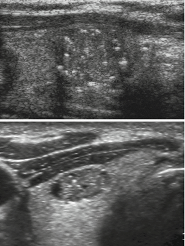
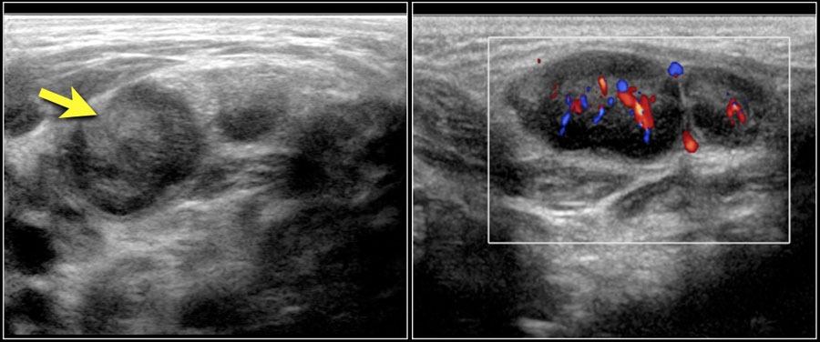
On color doppler it may show some peripheral vascularity in acute phase and may resemble breast cancer requiring a biopsy to establish tissue diagnosis. To document differences in color doppler flow and gray scale ultrasonographic us features between benign and malignant axillary lymph nodes in women with primary breast cancer. As lymph fluid leaves the breast and eventually goes back into the bloodstream the lymph nodes try to catch and trap cancer cells before they reach other parts of the body. The longitudinal transverse axis ratio and hilar status on color doppler flow and gray scale us images were prospectively studied for each of 145 axillary nodes in 135 women 74.
Axillary lymph node aln status is the most important prognostic factor for the management of breast cancer patients in the absence of metastatic disease. The analysis of patterns of nodal vascularity can be used to differentiate benign from malignant lymphadenopathy with high sensitivity. However fat necrosis usually exhibits no significant blood flow figure 16. Gray scale parameters that favor malignancy size.
The vascular flow is inadequately evaluated. A two dimensional color doppler sonography reveals rounded lymph node morphologic characteristics with loss of hilar fat. In 1998 reports that the use of color doppler sonography on axillary lymph nodes to diagnose the metastatic axillary lymph nodes of breast cancer. However the normally directed hilar blood flow arrow which is best seen with manipulation of the images supports the histologic diagnosis of a reactive lymph node.
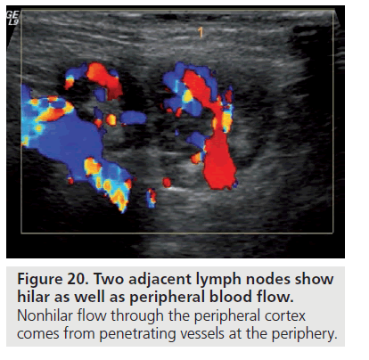
Color doppler ultrasound can show flow in all lymph nodes regardless of whether they are affected by a benign or a malignant process.
Breast Cancer Topic Lymph Nodes On Ultrasound
Intramammary Lymph Nodes Radiology Reference Article Radiopaedia Org

Lymph Node Matting Multiple Malignant Nodes Are Fused In A Single Ill Download Scientific Diagram

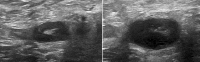
Nodal Echogenic Hilum A Benign Node With Echogenic Central Hilum B Download Scientific Diagram
Https Journals Sagepub Com Doi Pdf 10 1177 8756479305278268

A 75 Year Old Woman With Primary Breast Lymphoma Ultrasound Image Of Download Scientific Diagram

Benign Node Oval Shape Preserved Central Hilum Download Scientific Diagram

Axillary Lymph Nodes In Breast Cancer Patients Sonographic Evaluation

Diagnostic Efficacy Of Color Doppler Ultrasound In Evaluation Of Cervical Lymphadenopathy Abstract Europe Pmc

An Axillary Lymph Node Showing Indeterminate Morphology On Conventional Download Scientific Diagram

Ultrasound Scanning Of The Pelvis And Abdomen For Staging Of Gynecological Tumors A Review Fischerova 2011 Ultrasound In Obstetrics Amp Gynecology Wiley Online Library

Three Dimensional Sonography Of Axillary Lymph Nodes In Patients With Breast Cancer Koenigsberg 2016 Journal Of Ultrasound In Medicine Wiley Online Library

Pdf Mistakes In Ultrasound Diagnosis Of Superficial Lymph Nodes
Http Pdf Posterng Netkey At Download Index Php Module Get Pdf By Id Poster Id 112347

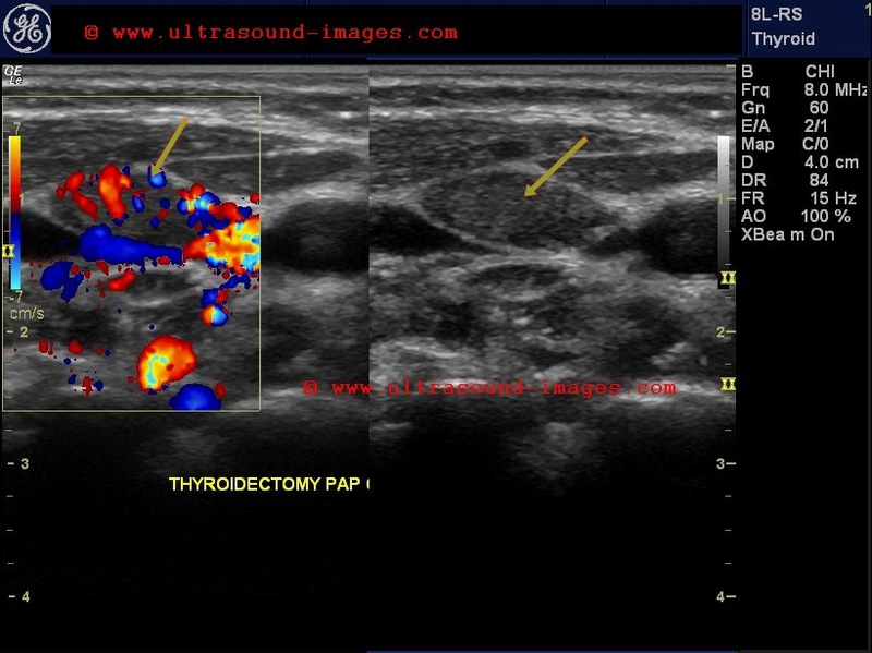
Pdf Diagnostic Approach For Evaluation Of Lymph Node Metastasis From Thyroid Cancer Using Ultrasound And Fine Needle Aspiration Biopsy
Ultrasound Features Of Extranodal Extension In The Metastatic Cervical Lymph Nodes Of Papillary Thyroid Cancer A Case Control Study Mu Cancer Biology Medicine

The Role Of Preoperative Ultrasound Evaluation Of Inguinal Lymph Nodes In Patients With Vulvar Malignancy Sciencedirect

Pin On Breast Imaging
Https Encrypted Tbn0 Gstatic Com Images Q Tbn 3aand9gcs7s1f45hgr7vullszgzhelfimsjzsoglyqns9f0efp104yj3pr Usqp Cau

Pdf Review Of Ultrasonography Of Malignant Neck Nodes Greyscale Doppler Contrast Enhancement And Elastography
Evaluation Of Neck Lymph Node Metastasis On Contrast Enhanced Ultrasound An Animal Study
Ultrasound Features Of Thyroid Parathyroid Neck Lymph Nodes Normal And Pathologic Pattern Oncohema Key
A Gallery Of High Resolution Ultrasound Color Doppler 3d Images Thyroid 2
Https Pubs Rsna Org Doi Pdf 10 1148 Rg 27si075502

The Radiology Assistant Ultrasound In Acute Abdomen In 2020 Ultrasound Diagnostic Medical Sonography Abdomen
The Radiology Assistant Neck Masses In Children

Ultrasound Of Malignant Cervical Lymph Nodes Abstract Europe Pmc

Fighting For My Family Blog It Ovarian Cyst Treatment Ovarian Cyst Cystic Teratoma
Http Pdf Posterng Netkey At Download Index Php Module Get Pdf By Id Poster Id 115518

Utility Of Ultrasound Elastography To Differentiate Benign From Malignant Cervical Lymph Nodes Kanagaraju V Rakshith A V Devanand B Rajakumar R J Med Ultrasound

Grades Of Hydronephrosis According To The Society Of Fetal Urology Sfu Ultrasound Sonography Fetal

Pdf Conventional Ultrasound For Lymph Node Evaluation Update 2013
Dr Ahmed Esawy Salivary Gland Ultrasound
The Roles Of Ultrasonography In The Management Of Axillary Node Metastases In Breast Cancer
Http Pdf Posterng Netkey At Download Index Php Module Get Pdf By Id Poster Id 110187

Google Image Result For Http Flylib Com Books 2 953 1 Html 2 44 20 20thyroid Lymph Nodes Tumor Sinusitis

Image Result For Cerebellar And Cisterna Magna On Ultrasound

Pin On Ob Gyn Ultrasound 101 Mod 2
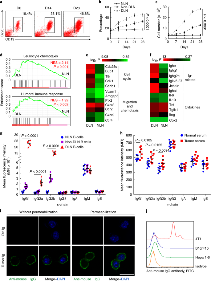
Tumor Educated B Cells Selectively Promote Breast Cancer Lymph Node Metastasis By Hspa4 Targeting Igg Nature Medicine

Pin On Does A Body Good

A Roadmap Of Ovarian Cyst Types Via Radiologyassistan

Ultrasonography Of Superficial Lymph Nodes Benign Vs Pages 1 13 Flip Pdf Download Fliphtml5

