Bpd Ultrasound Normal Range At 20 Weeks

Head Circumference Calculator Babymed Com

Estimation Of Fetal Weight Reference Range At 20 36 Weeks Gestation And Comparison With Actual Birth Weight Reference Range Salomon 2007 Ultrasound In Obstetrics Amp Gynecology Wiley Online Library

Biparietal Diameter Radiology Reference Article Radiopaedia Org

Fetal Ultrasound Biometry 1 Head Reference Values Kurmanavicius 1999 Bjog An International Journal Of Obstetrics Amp Gynaecology Wiley Online Library

Fetal Age Assessment Based On Femur Length At 10 25 Weeks Of Gestation And Reference Ranges For Femur Length To Head Circumference Ratios Johnsen 2005 Acta Obstetricia Et Gynecologica Scandinavica Wiley Online Library

Fetal Growth Between The First And Second Trimesters And The Risk Of Adverse Pregnancy Outcome Pedersen 2008 Ultrasound In Obstetrics Amp Gynecology Wiley Online Library

Different babies of the same weight can have different head size therefore dating in the later part of pregnancy is generally considered unreliable.
Bpd ultrasound normal range at 20 weeks. Your doctor is looking for the bpd measurement as well as the other measurements to be within what is considered normal range. The wide normal range of bpd in late pregnancy must be appreciated. Normal ultrasonic fetal growth ratios evaluated in cases of fetal disproportion. Biparietal diameter bpd this is the diameter between the two sides of the head and is measured after 13 weeks.
Ultrasound obstet gynecol 1994 4 34 48 the patients fulfilled the following. Femur length hadlock formulas. The reduced accuracy of prediction of gestational age after 20 weeks must be appreciated. Bpd and ofd were measured from the outer borders of the skull and head circumference hc was.
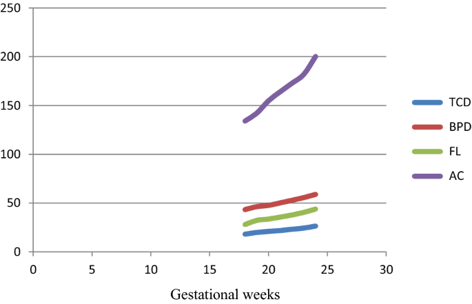
The use of the biparietal diameter bpd. The tcd curve was derived from 16 to 36 weeks of gestation. Biparietal diameter the diameter between the 2 sides of the head hc. Crang svalenius e jorgensen c.
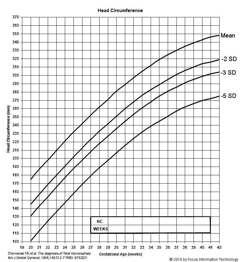
It increases from about 2 4 cm at 13 weeks to about 9 5 cm at term. Hohler cw quetel ta. Scroll down to see your guide to ultrasound. Ultrasound 18 20 weeks normal morphology scan head ultrasound the correct plane for the measurement of the head circumference hc and bi parietal diameter bpd must include the cavum septum pellucidum thallamus and choroid plexus in the atrium of the lateral ventricles.
Measurements within the normal range can also be assessed. This chart outlines expected ultrasound measurements in mm based on gestational age. Reference curves for normal fetal growth were developed from 10 weeks of gestation onwards for bpd hc and ac and from 12 weeks onwards for fl. The biparietal diameter measurement increases from roughly 2 4 centimeters at 13 weeks to approximately 9 5 centimeters when a fetus is at term.
J ultrasound med 1991 10 89 92. The aim of this study was to determine potential antenatal gender related differences in the ultrasound measurements of bpd hc ac and fl in uncomplicated singleton pregnancies the gestational age of which had been confirmed by early ultrasound. Am j obstet gynecol 1981 141 759 762. Itisrecommendedthatdepartments whichadoptthecharts tables in this document check that any data programmed into their ultrasound and computerised patient management sys tems use the ga and size equations given here.
If the ultrasound measurements are in agreement and differ from menstrual dates by more than one week prior to 20 weeks a new estimated due date should be calculated and recorded. Early ultrasound dating of pregnancy and the use of reliable growth curves can improve obstetric management in pregnancy.

Normal Cns Ultrasound Brain Anatomy Brain Anatomy Ultrasound Cns

Pin Em Obg

Pin On Fruits Of Our Labours

Ultrasound Image Of Dolicocephaly Diagnostic Medical Sonography Ultrasound Ultrasound Tech

Miscarriage After A Normal Scan At 12 14 Gestational Weeks In Women At Low Risk Of Carrying A Fetus With Chromosomal Anomaly According To Nuchal Translucency Screening Westin 2007 Ultrasound

Level Ii Usg
Microcephaly

Do I Keep Worrying 20 Week Ultrasound And Son S Head Is Measuring Just A Hair Under Normal Babycenter

Fetal Transverse Cerebellar Diameter Measurements In Normal And Reduced Fetal Growth Vinkesteijn 2000 Ultrasound In Obstetrics Amp Gynecology Wiley Online Library

Biparietal Diameter At 11 13 Weeks Gestation In Fetuses With Open Spina Bifida Khalil 2013 Ultrasound In Obstetrics Amp Gynecology Wiley Online Library

Isuog Practice Guidelines Performance Of First Trimester Fetal Ultrasound Scan 2013 Ultrasound In Obstetrics Amp Gynecology Wiley Online Library

Fractional Fetal Thigh Volume In The Prediction Of Normal And Abnormal Fetal Growth During The Third Trimester Of Pregnancy Sciencedirect

Small Biparietal Diameter In Fetuses With Spina Bifida On 11 13 Week And Mid Gestation Ultrasound Karl 2012 Ultrasound In Obstetrics Amp Gynecology Wiley Online Library

Fetal Biometry In Ethnic Chinese Biparietal Diameter Head Circumference Abdominal Circumference And Femur Length Leung 2008 Ultrasound In Obstetrics Amp Gynecology Wiley Online Library

Normal Ranges For Fetal Femur And Humerus Diaphysis Length During The Second Trimester In An Iranian Population Tahmasebpour 2012 Journal Of Ultrasound In Medicine Wiley Online Library

Fetal Size Monitoring And Birth Weight Prediction A New Population Based Approach Gjessing 2017 Ultrasound In Obstetrics Amp Gynecology Wiley Online Library

Fetal Ultrasound Biometry 2 Abdomen And Femur Length Reference Values Kurmanavicius 1999 Bjog An International Journal Of Obstetrics Amp Gynaecology Wiley Online Library

Pin On Ultrasound
Https Encrypted Tbn0 Gstatic Com Images Q Tbn 3aand9gct55r2fjxzolarplu6wj21vtlxvy5lws7snf7ci0pcrgunjvkkz Usqp Cau

Normal And Abnormal Development Of The Fetal Anterior Fontanelle A Three Dimensional Ultrasound Study Paladini 2008 Ultrasound In Obstetrics Amp Gynecology Wiley Online Library

Fetal Growth Reference Ranges In Twin Pregnancy Analysis Of The Southwest Thames Obstetric Research Collaborative Stork Multiple Pregnancy Cohort Stirrup 2015 Ultrasound In Obstetrics Amp Gynecology Wiley Online Library
Https Onlinelibrary Wiley Com Doi Pdf 10 7863 Jum 1984 3 5 227

Ultrasound Estimation Of Birth Weight In Twin Pregnancy Comparison Of Biometry Algorithms In The Stork Multiple Pregnancy Cohort Khalil 2014 Ultrasound In Obstetrics Amp Gynecology Wiley Online Library
Determination Of Fetal Transcerebellar Diameter Nomogram In The Second Trimester Springerlink
Https Onlinelibrary Wiley Com Doi Pdf 10 7863 Jum 1991 10 2 89

Fetal Sex And Intrauterine Growth Patterns Melamed 2013 Journal Of Ultrasound In Medicine Wiley Online Library

A Thai Reference For Normal Fetal Nasal Bone Length At 15 To 23 Weeks Gestation Sutthibenjakul 2009 Journal Of Ultrasound In Medicine Wiley Online Library

International Journal Of Current Research And Review Ijcrr

Normal Cns Ultrasound Brain Anatomy There Are Three Major Scan Planes For The Fetal Brain Which Accommodate 3 Views 1 Thalami Brain Anatomy Cns Ultrasound
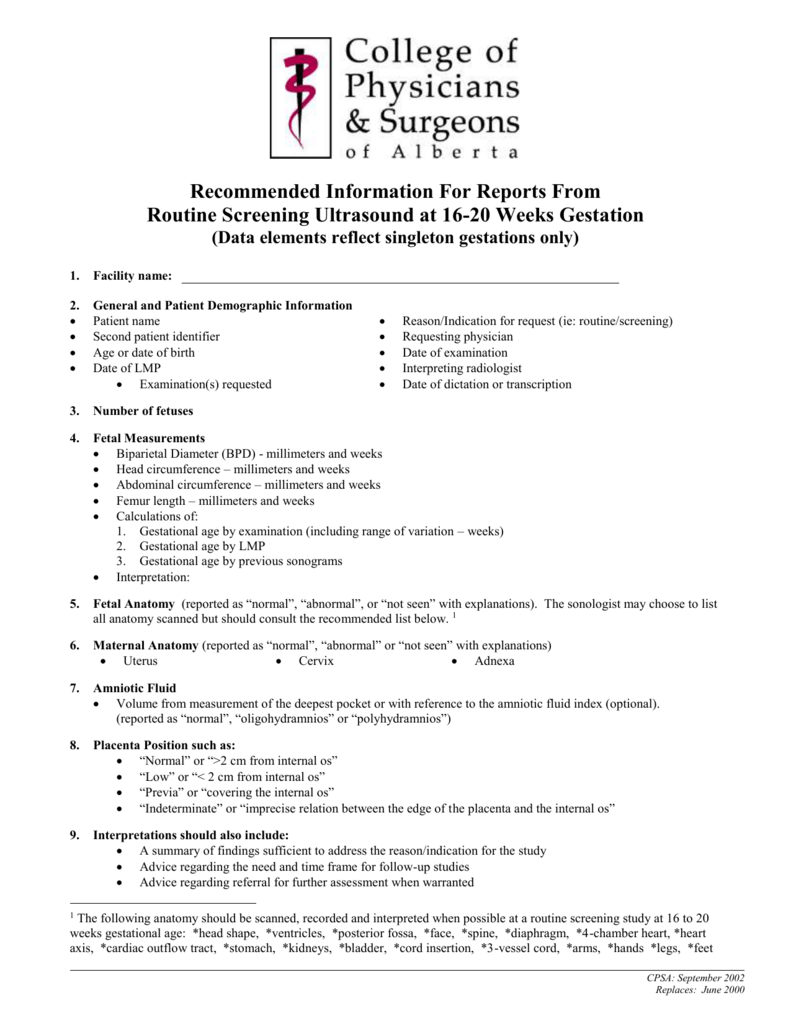
Routine Screening Ultrasound Recommendations
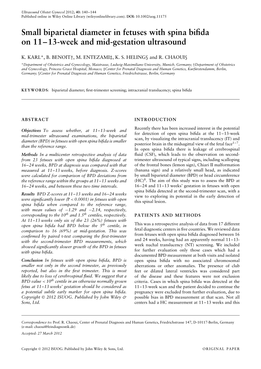
Pdf Small Biparietal Diameter In Fetuses With Spina Bifida On 11 13 Week And Mid Gestation Ultrasound
Depth Of Brain Fissures In Normal Fetuses By Prenatal Ultrasound Between 19 And 30 Weeks Of Gestation Alonso 2010 Ultrasound In Obstetrics Amp Gynecology Wiley Online Library

The Use Of Obstetric Ultrasound In The Antenatal Diagnosis Of Craniosynostosis We Need To Do Better Constantine 2016 Australasian Journal Of Ultrasound In Medicine Wiley Online Library
Https Onlinelibrary Wiley Com Doi Pdf 10 7863 Jum 1994 13 12 937

Pdf Normal Ranges Of Biorbital And Interorbital Distances In Healthy Turkish Pregnancies At 19 23 Weeks Of Gestation And Correlation With Craniofacial Structures
Https Journals Sagepub Com Doi Pdf 10 1177 8756479316687997

Intrauterine Growth Restriction Identification And Management American Family Physician

Pdf Serial Head Ultrasound Studies In Preterm Infants How Many Normal Studies Does One Infant Need To Exclude Significant Abnormalities
Pdf Prediction Of Newborn Birth Weight Based On The Estimation At 20 24 Weeks Of Gestation
Week 32 Ultrasound What It Would Look Like Parents

Normal Values In Pregnancy Content Last Reviewed 20th February 2020 Chapter 74 High Risk Pregnancy

